Figure 2.
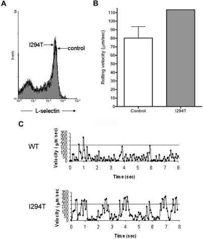
WASpI294T PBMCs demonstrate defective L-selectin–dependent rolling despite normal levels of surface L-selectin. (A) WT and WASpI294T PBMCs were freshly isolated from whole blood and monitored for surface L-selectin expression by flow cytometry. Histogram gated on PBMCs reveals comparable levels of L-selectin between patient and control samples. (B) A fixed density of patient or control PBMCs (ie, 3 × 10⁵/mL) were perfused over immobilized sLex at 2.5 dyn/cm2. A single patient sample was analyzed, whereas 3 control samples were analyzed; the average of 3 independent experiments is represented in the histogram. Approximately 30 cells were analyzed per field of view, and an average of 100 cells was analyzed per experiment. Error bar represents SD. (C) Representative example of the jerky nature of PBMC rolling from control and patient samples. Individual cells were tracked for a total of 8 seconds. The superimposed line represents a velocity of 200 μm/s, which is the velocity threshold above which cells are normally considered to be in free flow.

