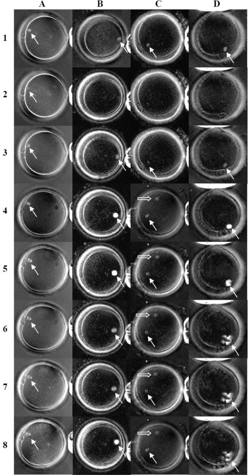
Figure 2.
Spindle images of human oocytes following cooling and treatment with cryoprotective agents (Experiment 1). Images in column A, B, C, and D (representing four treatment groups, PROH, EG, DMSO and taxol respectively) were taken after oocytes were maintained in PBS at 37°C (row 1), after temperature dropped to 20°C (row 2), then rewarmed to 37°C (row 3), after having been equilibrated with the cryoprotective agents at 37°C (row 4), after temperature dropped to 20°C (row 5), 10°C (row 6), 0°C (row 7) and then rewarmed to 37°C (row 8). Arrow outlines show spindles while solid arrows in column C indicate newly formed extra spindle structures [original magnification = 200X].

