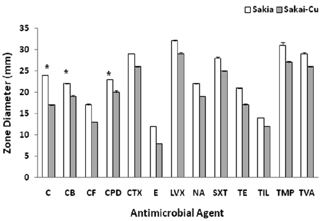
Figure 3.
Comparison of Sakai and Sakai-Cu for antibiotic susceptibility. Sakai and Sakai-Cu were streaked on Muller-Hinton agar and the antibiotic disks were placed on the plate. After incubation at 37°C for 18 h, the diameter of the growth inhibition zone was measured. The full names of the antibiotics are described in the text. Asterisks indicate differences from susceptible to intermediate (C and CPD) or intermediate to resistant (CB).

