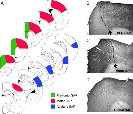
Figure 4.
Histological analysis of site-specific cholinergic lesions. (A) Series of line drawings demonstrating the extent of cholinergic depletion following focal intraparenchymal injections of the immunotoxin SAP into either the PFC, motor cortex, or AUD. Quantitative analysis of the extent of loss in innervation is presented in Table 1. Site-specific lesions depleted cholinergic inputs to the targeted region, creating a well-defined demarcation with adjacent cortical structures. For instance, injections of the immunotoxin into the dorsolateral PFC resulted in near complete loss of cholinergic fibers from within the prelimbic and cingulated cortices but did not affect innervation of the adjacent motor cortex (B). Following focal injections of the immunotoxin into motor cortex, a sharp demarcation was observed between the depleted motor cortex and the unaffected PFC (C). Global cholinergic lesions, generated by injecting the immunotoxin directly within the nucleus basalis/substantia innominata, depleted cholinergic innervation to both the prefrontal and motor cortices (D). Panels B–D were taken at the level indicated by the box in the line drawings in panel (A).

