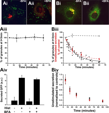
Figure 5.
The tPA-EGFP organelle is a short-lived compartment from which unstimulated secretion of tPA-EGFP and cytokines arises. (A-B) Representative examples of fixed cells expressing proregion-EGFP (Ai-ii; 24 hours after Nucleofection) or tPA-EGFP (Bi-ii; 6-7 hours after Nucleofection) and stained with a specific antibody to TGN46 (red in color images) in the absence of BFA (vehicle control; Ai and Bi) or after 1-hour treatment with 5μM BFA. Solid squares and line in panel Aiii represent the mean numbers of WPBs, expressed as a percentage of the total number at t = 3 minutes after addition of BFA, present in cells at the times indicated (n = 6 cells; BFA added at t = 0 minutes). Numbers of WPBs in vehicle-treated control cells at 3, 15, and 60 minute time points, expressed in the same way, are shown in open squares with dashed line (n = 4 cells). (Biii) Similar data for tPA-EGFP-containing puncta. In this case, the numbers of tPA-EGFP-containing granules in BFA-treated cells are plotted individually because of differences in the precise timing of measurements between experiments (n = 9 cells). The solid black line indicates a single exponential decline fitted to the pooled data for all cells. Superimposed on the plot (red circles and red broken line) is the mean decrease in unstimulated secretion of tPA-EGFP in cells treated with BFA. These data are expressed as a percentage of control cells (vehicle-treated) at the times indicated (n = 4 independent experiments each carried out in duplicate; BFA added at t = 0 minutes). Panels Aiv represent ELISA data for secreted EGFP from cells expressing proregion-EGFP. Cells exposed to BFA or vehicle control (1 hour) were stimulated with histamine or vehicle control (10 minutes) as indicated. Data shown are an individual experiment carried out in triplicate and are representative of 3 separate experiments. Panel Biv represents the mean decrease in unstimulated secretion of IL-6 (□), GRO-α (■), and MCP-1 (●) from IL-1β–treated cells exposed to BFA at t = 0. Data are normalized to that of vehicle control-treated cells at the times indicated and represent data pooled from 3 or 4 independent experiments each carried out in duplicate. For comparison, the data for unstimulated secretion of tPA-EGFP in BFA treated-cells plotted in panel Biii are included in red.

