Figure 6.
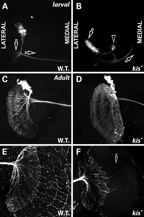
Kismet is required for proper DC position and axon migration in developing DCNs. All panels show GFP in DCNs by MARCM analysis. (A and B) Late third instar larval brains. Both images show the left hemisphere. Anterior is up. Medial is right. Lateral is left. (C–F) Adult brains 48 h after eclosion. (A) Wild-type MARCM clones (FRT 40A) in larval DCNs. Note position of the soma cluster axon bundles (parallel to vertical arrow) compared with commissural axon bundles (below horizontal arrow). (B) kisLM27 homozygous mutant MARCM clones display abnormal positioning of soma cluster compared with commissural axon bundles (left arrow), soma that have developed outside of the normal cluster (arrowhead) and disrupted commissural axon migration (right arrow). (C) Wild-type MARCM clones (FRT 40A) in adult DCNs displaying normal dendritic morphology on ipsilateral brain hemisphere. (D) kisLM27 homozygous mutant MARCM clones in adult brains. Overall morphology of dendritic field appears normal. (E) Wild-type MARCM clones (FRT 40A) in adult DCNs displaying normal axonal morphology on contralateral brain hemisphere. (F) kisLM27 homozygous mutant MARCM clones in adult brains. Arrow displays abnormal and reduced number of axonal extensions from lobulla into the medulla.

