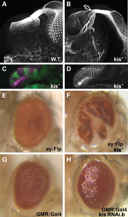
Figure 7.
Kismet is required for proper photoreceptor axon migration and eye development. (A–D) Late third instar larval retinas with attached brains. (E–H) Adult eyes, anterior right. (A) Wild-type retina with attached larval brain stained for the Futsch protein (white) display normal axonal bundles in the optic stalk that innervate the developing optic lobes. (B) kisLM27 homozygous mutant clones in the developing retina stained for Futsch (white) display abnormal axonal migration into the developing brain. Arrow indicates defasciculation of axon bundles in the optic stalk. (C) Cross-section of kisLM27 homozygous mutant clones in the developing retina posterior to the morphogenetic furrow. Heterozygous (control) tissue is denoted by the presence of GFP (green). Homozygous clones are denoted by lack of GFP. Retinas are stained with Futsch (magenta). Note normal positioning of ommatidia clusters near the apical (top) portion of the retina. (D) Futsch stain (white) from (C). (E) Normal eye expressing only eyeless:Flip. (F) Adult eye showing kisLM27 homozygous mutant clones (in white). (G) GMR-Gal4 adult eye raised at 25°C displays normal morphology. (H) GMR-Gal4 / UAS:kis RNAi.b adult eye raised at 25°C displays abnormal morphology and glassy appearance.

