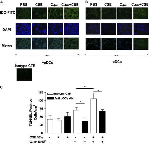
Figure 7.
The depletion of pDCs significantly decreased IDO and apoptotic cells in the lung. Mice were adoptively transferred with CSE-pulsed and/or C. pneumoniae–pulsed BMDCs, and lung sections were stained for IDO expression and TUNEL assay. The expression of IDO was determined by means of an immunofluorescence technique, using FITC either in the presence (A) or after the depletion (B) of pDCs. The isotype control (rat IgG2b) was used as negative control. (C) A TUNEL assay was performed, and positive cells were quantified. Data represent mean ± SEM, and each experiment was performed at least five times for each type of experimental condition. Statistically significant differences are denoted as *P < 0.05, as determined by one-way ANOVA and the Student t test.

