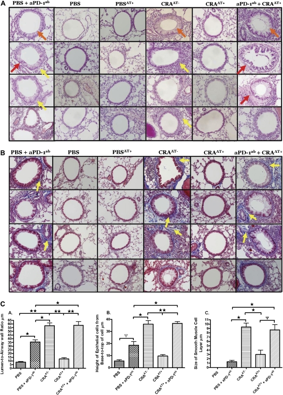
Figure 4.
Lung histology after adoptive transfer. (A) Lung sections obtained after 4 wk of cockroach aerosol challenges from each treatment group. Sections were stained with hematoxylin and eosin, and morphology was examined by light microscopy. Yellow arrows indicate inflammatory cell infiltration surrounding the airways. Orange arrows indicate hypertrophy of airway epithelial cells. Red arrows indicate smooth muscle cell hyperplasia and hypertrophy. (B) Lungs were removed, embedded with paraffin, and sectioned (5 μm) after 4 wk of cockroach aerosol and methacholine challenges from each treatment group after adoptive transfer. Sections were processed by trichrome staining and evaluated for collagen deposition by light microscopy, as indicated by yellow arrows. These data are representative of five mice per group. (C) These datasets provide a comparative analysis of each experimental group, quantifying morphometric changes in the airways of normal-to-induced asthmatic mice and treated mice. The morphometric parameters include (A) lumen-to-airway wall ratio, (B) epithelial cell height, and (C) changes in thickness of smooth muscle cell layer.

