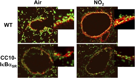
Figure 1.
Localization of NF-κB activation in lung sections from wild-type and CC10-IκBαSR transgenic mice exposed to NO2. The CC10-IκBαSR and wild-type (WT) littermate control animals were exposed to room air or 25 ppm NO2 for 6 hours, and killed 1 hour later. Lung sections were evaluated for nuclear translocation of RelA, using immunofluorescence and confocal laser scanning microscopy. Nuclei were visualized with Sytox (green), and RelA was visualized using a Cy3-conjugated secondary antibody (red). Nuclear localization is indicated by the overlap of fluorophores (yellow). Original magnification of large images is ×200, and is representative of patterns observed in at least four mice per group. For a better illustration of the differences in nuclear NF-κB localization, airway epithelial cells from the original images were magnified using a 2.5 optical zoom and are shown at upper right (insets).

