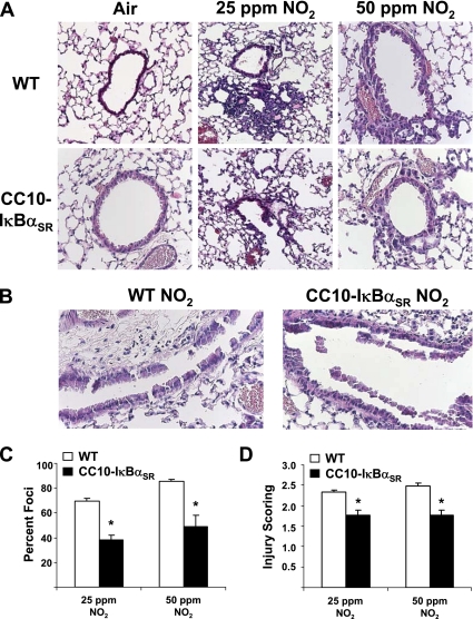
Figure 4.
Evaluation of histopathology in wild-type (WT) and CC10-IκBαSR mice exposed for 6 hours/day to 25 ppm or 50 ppm of NO2 for 3 days. (A) Hematoxylin-and-eosin staining of 5-μm sections of mouse lungs. (B) Hematoxylin-and-eosin staining of large airways from mice exposed to 50 ppm NO2. All magnifications are ×200, and are representative of patterns observed in each group. Scoring of lesions by assessment of the percentage of airways involved per section (C) and the intensity of foci (D) was performed as described in Materials and Methods (*P < 0.05 compared with WT mice; 25 ppm NO2, n = 8 mice/group; 50 ppm NO2, n = 5 mice/group).

