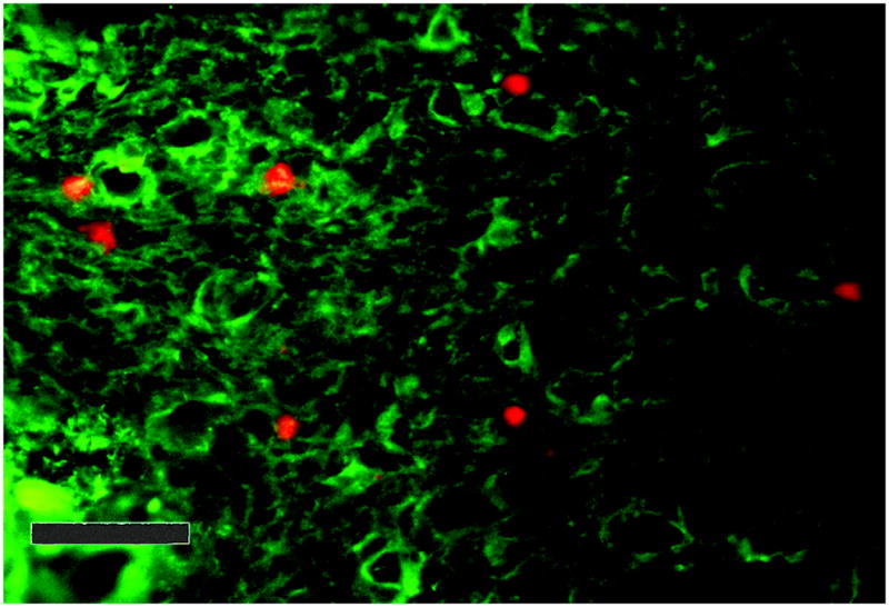
Figure 1.

Photomicrograph of results of Stamper-Woodruff adhesion assay. Human lymphocytes (5×10⁶ in 100 μL) were pre-labeled with anti-CD56-PE, then applied to decidua from a C57Bl/6J mouse at day 7 of gestation under rotation, to mimic shear forces in blood vessels. Murine endothelium was labeled with Alexa 488 conjugated isolectin (bright green). Non-adherent cells were rinsed off, the tissues were fixed and mounted. The number of CD56+ cells (bright red cells) per high power field (HPF) were counted. (400X). Size bar shows 50μm.
