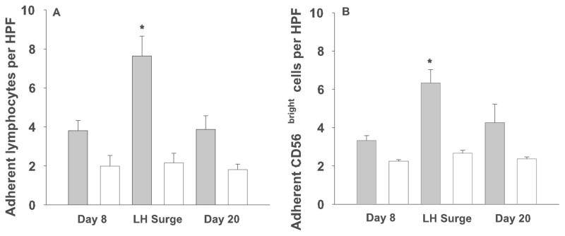
Figure 3.
Adhesion of lymphocytes from 3 timepoints in the menstrual cycle to LN and uterus from pregnant B6 mice.
Solid bars represent the mean number of adherent lymphocytes ± se (A) or CD56+ cells (B) per HPF in decidua from gd 6–8 B6 mice. Open bars show adhesion of lymphocytes pre-treated with a function-blocking antibody to L-selectin (n=6) which was significantly less than the untreated cells (p=0.0004, mixed linear model, F-test). * denotes significant increase in adhesion of untreated cells at LH as compared to untreated cells at cycle day 8 and 20 as determined by a mixed linear model, p=0.0004 (A) and p=0.0092 (B).

