
Figure 4.
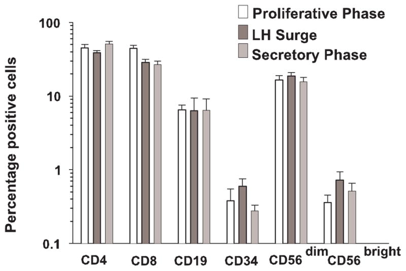
The relative percentages of lymphocyte subsets over the course of a menstrual cycle. As analyzed by flow cytometry, the numbers of CD4, CD8, CD19, CD33 and CD56dim cells remained stable. Each bar represents the mean of 3–6 samples.
Official websites use .gov
A
.gov website belongs to an official
government organization in the United States.
Secure .gov websites use HTTPS
A lock (
) or https:// means you've safely
connected to the .gov website. Share sensitive
information only on official, secure websites.
The relative percentages of lymphocyte subsets over the course of a menstrual cycle. As analyzed by flow cytometry, the numbers of CD4, CD8, CD19, CD33 and CD56dim cells remained stable. Each bar represents the mean of 3–6 samples.