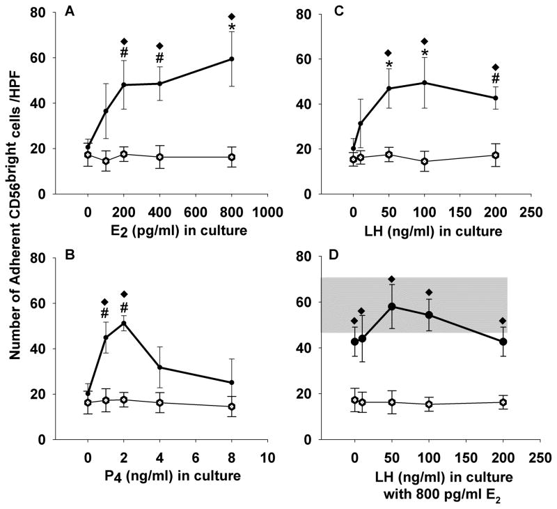
Figure 5.
Adhesion of cultured lymphocytes of male donors (n=4) to uterine endothelium. Peripheral blood lymphocytes were cultured with concentrations of 17β estradiol varying from 0 to 800 pg/mL (A), progesterone varying from 0 to 8 ng/mL (B), luteinizing hormone varying from 0 to 200 ng/mL (C) or luteinizing hormone varying from 0 to 200 ng/mL with 800 pg/ml 17β estradiol (D) for 4 h prior to assay for adhesion to frozen sections of mouse uterus. Gray bar in panel D shows effect of 800 pg/mL alone on adhesion (mean ± sem), without added LH. Data points represent mean values ± se. Open circles denote mean number of adherent CD56+ cells pre-treated with anti-L-selectin, closed circles show mean adhesion of untreated NK cells.* increased adhesion relative to control sample with no added hormones, p<0.005 (one way ANOVA), # increased adhesion relative to control sample with no added hormones, p<0.05 (one way ANOVA), ◆ differs from samples pre-treated with antibody to L-selectin p<0.008 (one way ANOVA).

