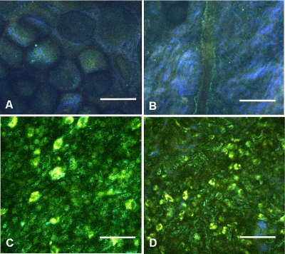
Figure 10.
NLOM images of rabbit synovial tissue demonstrating the enhanced image quality achieved by combining images acquired by sequential 730-nm and then 800-nm excitation. The images illustrated were obtained from the subintima in a plane parallel to and at a depth of 45 μm from the surface of (A) and (B) normal, (C) 8 days postbacterial inoculation, and (D) 18 days post-LPS intra-articular injection specimens. At a depth of 45 μm, the imaging plane is below the intimal layer of the normal synovium and demonstrates subintimal tissue predominately consisting of (A) adipocytes or (B) collagen matrix with little other cellular content, while both the (C) septic and (D) LPS specimens demonstrate a substantial increase in cellular content consistent with the inflammatory processes. The septic tissue demonstrates large cells with multilobular nuclei characteristic of rabbit heterophils and macrophages, along with elongated cells characteristic of fibroblasts, while the cellular infiltrate observed in the LPS specimens are smaller cells with oval nuclei characteristic of an inflammatory response predominated by lymphocytes. SHG emission of collagen, viewed as blue fibrils in the NLOM images, is not observed in the subintimal tissue of the septic specimens in (C), while it continues to be observed, although in reduced amounts, in LPS specimens in (D). Scale bars are 50 μm. (Color online only.)

