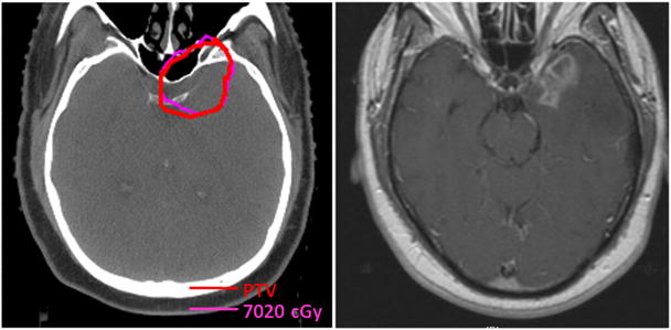
Fig 5.

Axial CT*-based planning slice (left) through the PTV† of a patient with intracranial tumor extension who developed temporal lobe necrosis (MRI‡ right) in a region that received prescription dose.
Abbreviations: * = Computed tomography; † = Planning target volume; ‡ = Magnetic resonance imaging.
