
Fig. 2.
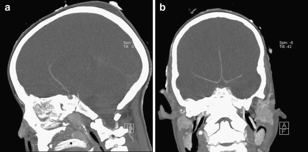
CTA images of a 7-year-old girl with brain death according to clinical criteria. Six-millimeter maximum intensity projection in coronal (a) and sagital (b) plane of CTA images obtained 60 sec after contrast material injection. The pericallosal arteries and the basialar artery are opacified. The internal cerebral veins, the great cerebral vein, and the straight sinus are not opacified. Based on the criteria, brain death cannot be confirmed