
Figure 1.
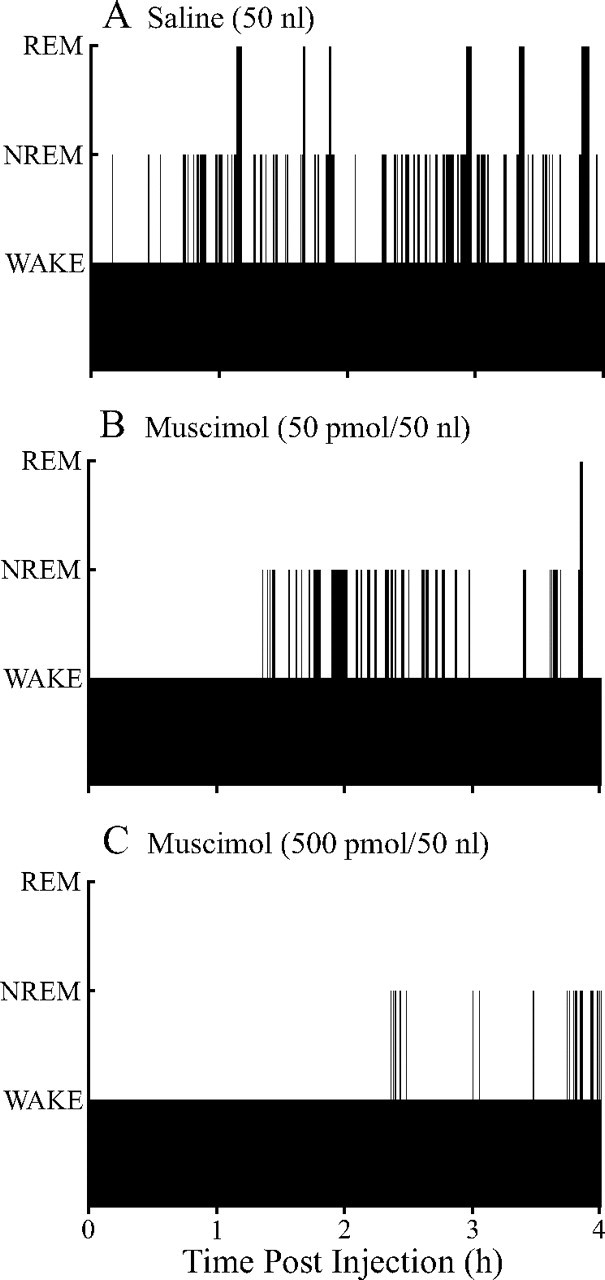
Microinjection of muscimol into the PnO alters the distribution of sleep and wakefulness. Black bars plot the time course of sleep and wakefulness recorded from the same mouse for 4 h after microinjection of saline (vehicle control) (A) and two concentrations of muscimol (B, C). The height of the bars corresponds to arousal state, with the lowest bars indicating wakefulness, intermediate bars representing NREM sleep, and highest bars showing the occurrence of REM sleep. Latency to onset of the first episode of NREM sleep and REM sleep was measured from time 0, which marks the end of the 1 min period during which saline or muscimol was delivered to the PnO.