
FIG. 3.
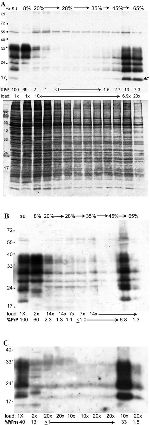
Sonicated supernatant samples loaded on more concentrated sucrose steps were centrifuged for a reduced time of 1.25 h (259,000 g-h). The top gradient (A) was loaded with 5 × 10⁷ cell equivalents, and the PrP Western blot with corresponding gold stain are shown. Unlike the 2.5-h gradients in Fig. 2, substantially more PrP (69%) remains at the top of the gradient (8% sucrose layer); 97% of the loaded PrP was recovered from this gradient. Even without proteinase K digestion, smaller PrP-res bands (as at arrow) are more prominent in the lower two gradient fractions that together contain 20% of the loaded PrP, as well as reduced protein by gold staining. Gradients B and C show PrP and PrP-res in a repeat gradient loaded with 108 cell equivalents in which the relative %PrP-res was rigorously quantified. Panel B shows the undigested PrP samples, and panel C shows aliquots of the same samples digested with proteinase K. Again, the majority of total PrP remains at the top of the gradient, with less than 10% in the bottom two fractions. Lane loads and the percentage of PrP and PrP-res in each fraction are indicated. In each of the gradients ~7 mL of sonicated supernatant in 8% sucrose was loaded on top of a step gradient made with (from top to bottom) 1 mL, 2 mL, 1 mL, 1 mL, and 0.3 mL, respectively, of the sucrose concentrations indicated. As in previous gradients, the entire supernatant was collected as a single fraction (up to the interface), and ~0.5-mL fractions collected from the 20% sucrose step downwards.