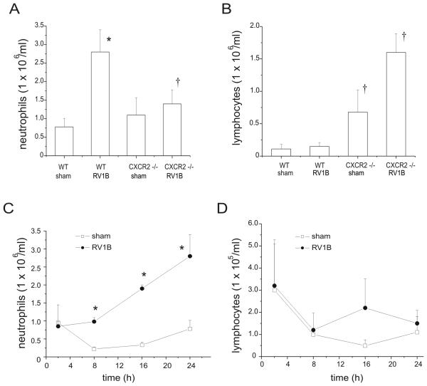
Figure 3.
Effects of CXCR2 knockout on lung inflammatory cells in sham and RV-treated mice. Mouse lungs were isolated, minced, and then digested in collagenase type IV for 1 h. Leukocytes were enriched following RBC lysis treatment, and counted for the presence of neutrophils (A, C) and lymphocytes (B, D). Panels A and B show data from 24 h after infection. Panels C and D show the early time course of neutrophil and lymphocyte influx in wild type mice. (N = 5 mice per group, bars represent mean±SEM, *different from respective sham group, p<0.05, ANOVA; †different from RV1B-treated wild- type mice, p<0.05, one-way ANOVA.)

