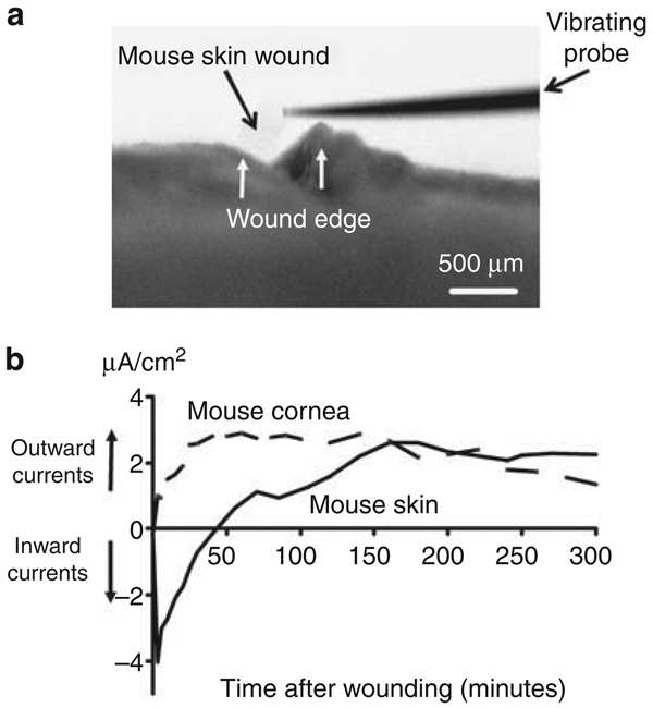
Figure 1. Endogenous electric currents at skin wounds.
(a) View down dissecting microscope showing a vibrating probe in measuring position at a shallow lancet wound on the skin of a mouse. Bar = 500 µm. (b) Time courses of endogenous electric currents. Note the slow rise and long-lasting nature of the skin wound electric currents. Measurements were carried out immediately after cervical dislocation and wounding. Positive value: outward current; negative value: inward current. The data are grouped from three independent experiments with three individual mice.

