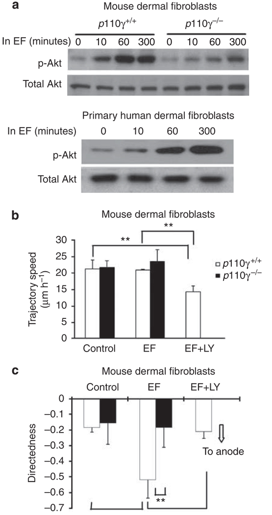
Figure 6. Role of phosphatidylinositol-3-OH kinase (PI3 kinase) signaling in electric field (EF)-directed migration of dermal fibroblast (DF) cells.
(a) Western blot analysis shows that exposure to an EF increased cell levels of phosphorylated Akt (p-Akt), indicating activation of PI3 kinase signaling. Knockout of PI3 kinase catalytic subunit (p110γ) significantly reduced PI3 kinase signaling. (b, c) Knockout of p110γ abolished directional migration, whereas the migration speed did not change significantly. Pharmacological inhibition of PI3 kinase also resulted in loss of directional migration in an EF. Cell numbers were 59–69, and 60–92 for p110γ+/+ and p110γ−/− cells, respectively, from three independent experiments. EF = 100 mV mm−1. **P<0.01.

