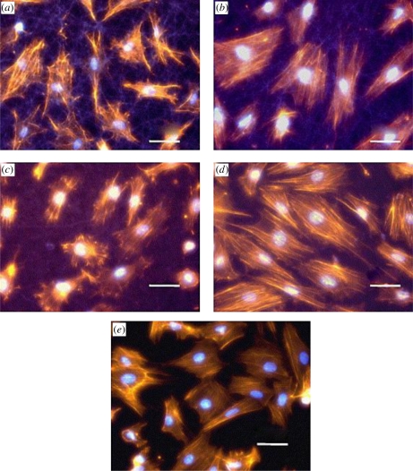
Figure 14.
Morphology of H9c2 myoblast cells at 20 h of post-seeding on: (a) gelatin fibre, (b) 15 : 85 PANI–gelatin blend fibre, (c) 30 : 70 PANI–gelatin blend fibre, (d) 45 : 55 PANI–gelatin blend fibres, and (e) glass matrices. Staining: nuclei, bisbenzimide; actin cytoskeleton, phalloidin; fibres, autofluorescence; original magnification 400× (Li et al. 2006). Scale bars, (a–e) 50 µm.

