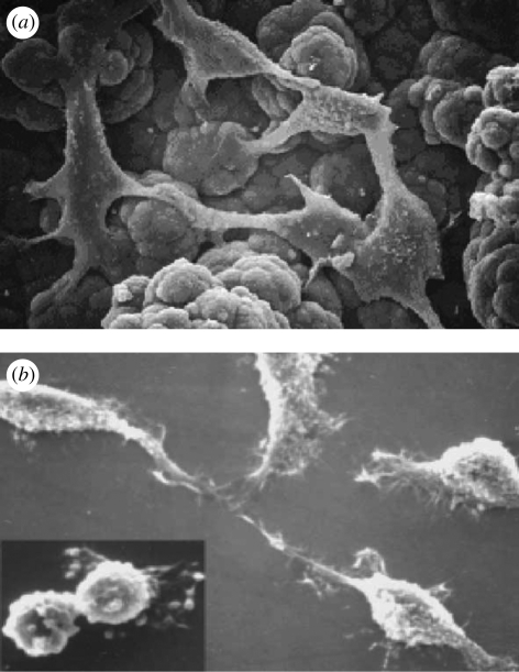
Figure 15.
SEM micrographs of NCTC keratinocytes (a) cultured onto self-standing oxidized PPy film (original magnification 2400 × ) and (b) grown onto thin oxidized PPy film showing different cell morphologies (original magnification 1000×). Inset: mitotic figure (original magnification 1500×; Mattioli-Belmonte et al. 2002).

