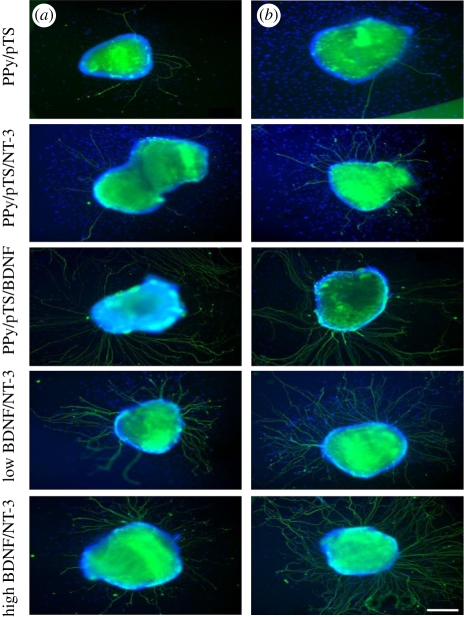
Figure 6.
Representative images of cochlear neural explants grown on PPy/pTS polymers with and without neurotrophin. Neurites were visualized by immunocytochemistry with a neurofilament-200 primary antibody and a fluorescent secondary antibody (green). Cell nuclei are labelled with DAPI (blue). (a) In the absence of neurotrophin (PPy/pTS), very few neurites were observed from explants, while explants grown on PPy/pTS containing neurotrophin demonstrated increased numbers of sprouting neurites. A greater number of neurites per explant was observed on explants grown on the electrically stimulated PPy films (b). These images were taken after 4 days of explant culture. Scale bar, 200 µm (Thompson et al. 2010).

