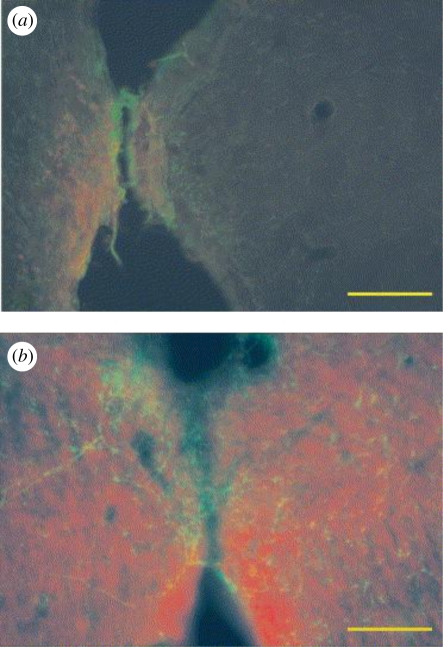
Figure 8.
A fluorescently labelled section of neural tissue in an implant lumen: (a) neural tissue in the lumen of the Teflon implants and (b) neural tissue in the PPy lumen where the glia has reformed and neurons are present; scale bar, 100 µm; green, glia; red, neurons (George et al. 2005).

