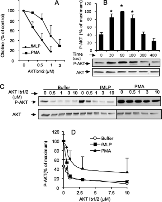
FIGURE 1.
PLD activity induced by fMLP or PMA in dHL-60 cells is abrogated by the AKT antagonist, AKTib1/2. A, PLD activity in dHL-60 cells pretreated without (control) or with 0.25–3 μm AKTib1/2 for 15 min before stimulation with 1 μm fMLP or 0.5 μm PMA for 3 min. The stimulated production of choline by PLD is expressed as a percentage of control values (n = 5 experiments). B, time course of AKT phosphorylation induced by 1 μm fMLP in dHL-60 cells and its densitometric quantification (*, p < 0.05). C, AKT phosphorylation in dHL-60 cells pretreated without (control) or with 0.5–10 μm AKTib1/2 for 15 min before cell stimulation with 1 μm fMLP for 1 min or 0.5 μm PMA for 3 min. A representative Western blot of phospho-AKT (Ser473) (P-AKT) is shown in C, and the densitometric quantification (D) is expressed as percentage of maximal response (n = 4 experiments).

