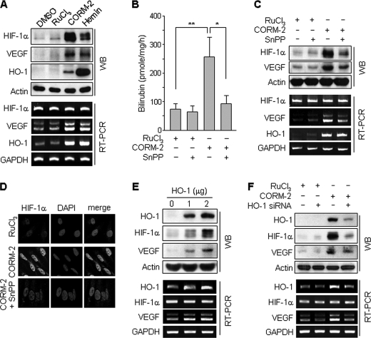
FIGURE 2.
CORM-2-induced HIF-1α and VEGF expressions are regulated by HO-1 activity. A, astrocytes were treated with DMSO, RuCl3 (200 μm), CORM-2 (100 μm), and hemin (10 μm) for 8 h. B, astrocytes were transfected with mock or HO-1 expression vector and cultured in fresh medium for 48 h. HO activity was assayed as described under “Experimental Procedures.” Data shown are the mean ± S.D. (n = 3). *, p < 0.05; **, p < 0.01. C, astrocytes were treated with RuCl3 (200 μm) or CORM-2 (100 μm) for 8 h in the presence or absence of SnPP (50 μm). The protein and mRNA levels of HIF-1α, VEGF, and HO-1 were analyzed by Western blotting and RT-PCR. D, cells treated with RuCl3 (200 μm), CORM-2 (100 μm), or CORM-2 plus SnPP (50 μm) for 8 h were immunostained with an antibody against HIF-1α (green) and DAPI (blue). Fluorescent images were obtained by a confocal microscope. E, astrocytes were transfected with mock or HO-1 expression vector and cultured in fresh medium for 48 h. F, astrocytes were transfected with control or HO-1 siRNA (50 nm), cultured in fresh medium for 40 h, and then treated with 200 μm RuCl3 or 100 μm CORM-2 for 8 h. E and F, the protein and mRNA levels of HIF-1α, VEGF, and HO-1 were analyzed by Western blotting and RT-PCR. Error bars, S.D. WB, Western blot.

