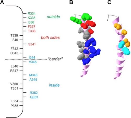
FIGURE 9.
Proposed location of internally exposed side chains in TM6. A, location of residues which, when mutated to cysteine, are exposed to internal MTS reagents only (blue), to external MTS reagents only (green), or to MTS reagents applied to either side of the membrane (red). Other resides that we find not to be modified by internal MTS reagents and presumably not exposed to the pore lumen are shown in black. Internal MTS modification is as defined in the present work; external MTS modification as shown in Fig. 6 (T338C, S341C) or in previous work (26, 28–31). Note that F337C has been identified as accessible to external MTS reagents in some previous studies (29, 31) but not in others (28, 30); it is included as pore-lining here because we find it to be accessible to internal MTS reagents (Fig. 2). B, shown is the location of these side chains in a recently published atomic homology model of TM6 (24), viewed from the side. Side chains allocated as accessible from the outside, accessible from the inside, accessible from both sides, or inaccessible, are colored as in A, whereas the TM6 helical backbone is colored pink. C, shown is the proposed location of state-dependent change in side chain accessibility in the same model of TM6. Side chains accessible only in activated channels (Thr-338, Ser-341, Ile-344) are colored orange, and those accessible both in activated and non-activated channels (Val-345, M348) are in cyan. Other side chains are not shown.

