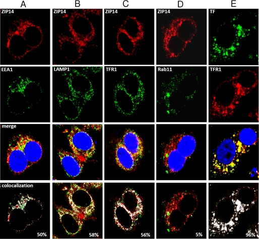
FIGURE 6.
Confocal microscopic analysis of the subcellular localization of ZIP14 in HepG2 cells. Fixed and permeabilized cells were analyzed for endogenous ZIP14–3×FLAG by using anti-FLAG antibody followed by rhodamine-conjugated secondary antibody. A–D, co-localization of ZIP14 with EEA1 (A), LAMP1 (B), TFR1 (C), and Rab11 (D) was determined by using respective primary antibodies followed by Alexa Fluor 488-labeled secondary antibody. Merged images show co-localization of ZIP14, with nuclei stained by DAPI. E, as a control, TF was co-localized with TFR1 by first incubating cells for 30 min with Alexa Fluor 488-labeled human holo-TF prior to fixation and permeabilization of cells. TFR1 was determined by using anti-TFR1 antibody followed by rhodamine-conjugated secondary antibody. All images were obtained by using a Leica TCS SP5 laser-scanning confocal microscope. Areas of co-localization (designated by white) were determined by using the co-localization tool provided with the Leica SP5 software.

