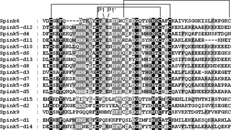
FIGURE 2.
Amino acid sequence alignment of SPINK6 in comparison with LEKTI/SPINK5 domains and SPINK6. The alignment of the Kazal domains of SPINK6, LEKTI-2/SPINK9, and LEKTI/SPINK5 domains (d) was generated by ClustalW and displayed using GeneDoc. Identical residues are in black boxes, whereas gray boxes indicate partially conserved residues. The darker the shading, the more the amino acids are conserved among the family members. The different LEKTI/LEKTI-2 domains are ordered by their homology to SPINK6. The P1 and P1′ sites are indicated.

