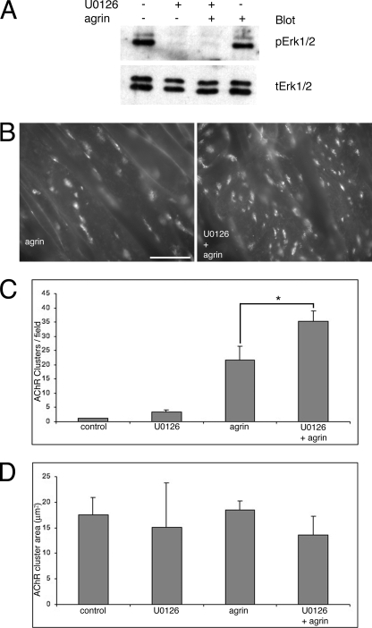
FIGURE 3.
Inhibition of ERK1/2 activation potentiates agrin-induced AChR clustering. A, Western blot for ERK1/2 at the end of a 1-h incubation with agrin, with or without U0126. The MEK inhibitor prevented ERK1/2 activation whether agrin was present or not. As shown in Fig. 1, levels of active ERK1/2 are not much different between untreated control and agrin-treated cells at 1 h after agrin addition. B, sample 400× fields of myotubes treated with agrin and agrin + U0126. There are many more AChR clusters (labeled with rhodamine-α-bungarotoxin) in the latter than the former. Scale bar, 50 μm. C, quantification of AChR clustering. There was ∼60% increase in AChR clusters in dishes treated with agrin + U0126 relative to dishes treated with agrin. Data are expressed as mean ± S.E. n = 4 for each treatment. *, p = 0.008, t test. D, quantification of AChR cluster area. No statistically significant differences were found in the AChR cluster area between any of the treatments. Data are expressed as mean + S.E. (error bars = + S.E.). n = 4 for each treatment.

