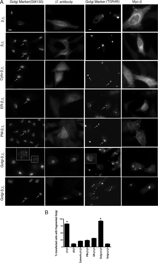
FIGURE 1.
Constitutively targeted Golgi-β1γ2 converts the Golgi into small vesicles. A, Myc-tagged β1 or β5 were expressed in HeLa cells along with wild-type or mutant γ2 in the following combinations, as indicated: Myc-β1 and γ2 (β1γ2), Myc-β5 and γ2 (β5γ2), Myc-β1 and Cyto-γ2 (Cyto-β1γ2), Myc-β1 and ER-γ2 (ER-β1γ2), Myc-β1 and PM-γ2 (PM-β1γ2), Myc-β1 and Golgi-γ2 (Golgi-β1γ2), and Myc-β5 and Golgi-γ2 (Golgi-β5γ2). Cells were visualized by immunofluorescence microcopy using anti-Myc, anti-GM130, or anti-TGN46 to detect β, the cis-Golgi stacks, and the trans-Golgi stacks, respectively. Arrows indicate intact Golgi. The inset shows vesiculated Golgi. Scale bar, 10 μm. B, results from A were quantitated and plotted as the percentage of cells with a fragmented Golgi. 100 cells from each of 6 different experiments were counted as having intact or fragmented Golgi. The percentage of cells with fragmented Golgi was >75% in β1γ2-expressing cells and >90% in Golgi-β1γ2-expressing cells, whereas in control transfections (empty vector) fragmented Golgi was observed in 10% of cells (not shown). Statistical significance as compared with control was tested using unpaired t test (*, p < 0.001).

