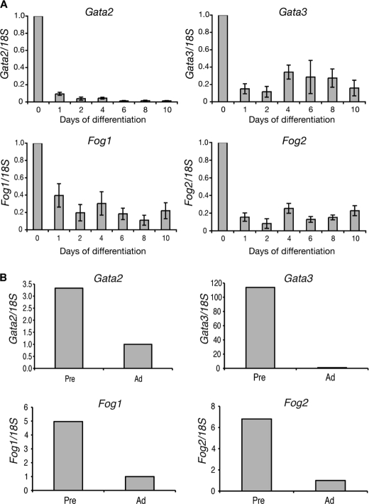
FIGURE 1.
Fog1 and Fog2 are expressed in 3T3-L1 cells and murine preadipocytes. A, real-time PCR analysis of Fog expression during adipogenesis. 3T3-L1 cells were induced to differentiate over 10 days. Samples were collected at the times indicated and analyzed. Expression of Gata2, Gata3, Fog1, and Fog2 was normalized with 18S and expressed relative to the Day 0 level. Error bars, S.E. (n = 3). B, epidydymal white adipose tissue from six 13-week-old male mice was pooled and digested with collagenase. Preadipocytes (Pre) and adipocytes (Ad) were separated by centrifugation and used for real-time PCR analysis. Expression of Gata2, Gata3, Fog1, and Fog2 were normalized with 18S and expressed relative to the level in adipocytes.

