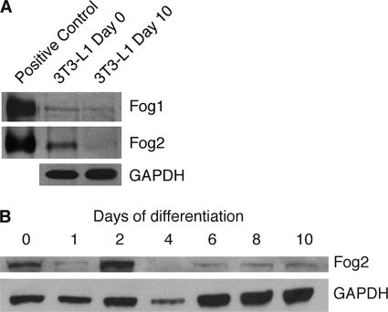
FIGURE 2.
FOG protein levels are reduced during adipogenesis. A, Western blot of 3T3-L1 cells at day 0 and day 10 of differentiation. Cells were harvested, and nuclear extracts were prepared. Positive controls of FOG1 and FOG2 overexpressed in HEK-239 cells were included. Western blotting was performed with FOG1 and FOG2 antibodies. Blotting with GAPDH antibody was included as a loading control. B, Western blot of 3T3-L1 cells at day 0, 1, 2, 4, 6, 8, and 10 of differentiation. Cells were harvested, and nuclear extracts were prepared. Western blotting was performed with a FOG2 antibody, and GAPDH antibody was included as a loading control.

