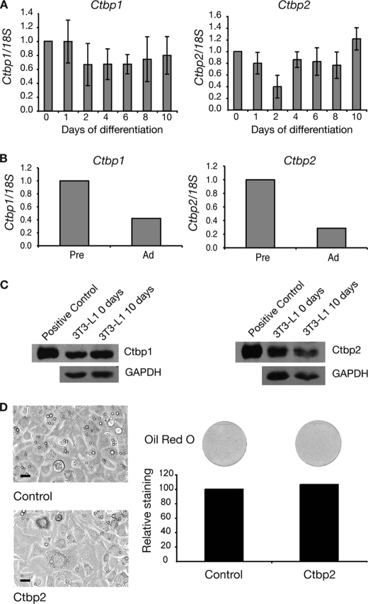
FIGURE 5.
Ctbp1 and Ctbp2 are expressed throughout 3T3-L1 differentiation. A, real-time PCR analysis of Ctbp expression during adipogenesis. 3T3-L1 cells were induced to differentiate over 10 days. Samples were collected at the times indicated and analyzed. Expression of Ctbp1 and Ctbp2 were normalized with 18S and expressed relative to the Day 0 level. Error bars, S.E. (n = 3). B, expression of Ctbp1 and Ctbp2 in mouse preadipocytes. Epididymal white adipose tissue from six 13-week-old male mice was pooled and digested with collagenase. Preadipocytes (Pre) and adipocytes (Ad) were separated by centrifugation and used for real-time PCR analysis. Expression of Ctbp1 and Ctbp2 was normalized with 18S and expressed relative to the level in adipocytes. C, Western blot of 3T3-L1 cells at day 0 and day 10 of differentiation. Cells were harvested, and nuclear extracts were prepared. Positive controls of CTBP1 and CTBP2 overexpressed in HEK-239 cells were included. Western blotting was performed with CTBP1 and CTBP2 antibodies. Blotting with GAPDH antibody was included as a loading control. C, images of 3T3-L1 cells expressing CTBP2 at 5 days of differentiation. Cells expressing CTBP2 or an empty vector control were induced to differentiate for 5 days. Cells were imaged by light microscopy. Scale bars, 20 μm. D, Oil Red O staining of 3T3-L1 cells expressing CTBP2 after 5 days of differentiation. Cells were stained with Oil Red O and imaged by scanning. The stain was extracted and quantified by spectrophotometry. Results are expressed as staining relative to control.

