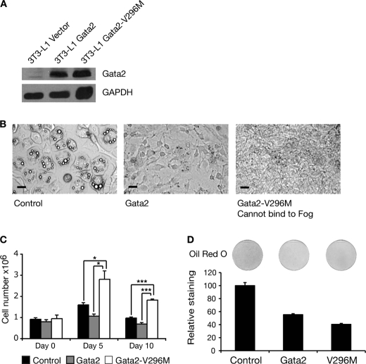
FIGURE 8.
Abolition of FOG binding activity in GATA2 impairs GATA2 function in 3T3-L1 cells. A, Western blot of GATA2 and GATA2-V296M overexpression in 3T3-L1 cells. Retroviral delivery was used to express GATA2 and GATA2-V296M in 3T3-L1 cells. The cells were grown to 2 days postconfluence, and nuclear extracts were prepared. Western blotting was performed with a GATA2 antibody. Blotting with GAPDH antibody was included as a loading control. B, images of 3T3-L1 cells expressing GATA2 and GATA2-V296M after 5 days of differentiation. Cells expressing GATA2, GATA2-V296M, or an empty vector control were induced to differentiate for 5 days. Cells were imaged using light microscopy. Scale bars, 20 μm. C, 3T3-L1 cells expressing GATA2, GATA2-V296M, or an empty vector control were induced to differentiate over 10 days. Cells were harvested at the indicated times, and the number of cells was determined by counting. Error bars, S.E. (n = 3). Student's t test results are shown as follows: *, p < 0.05; **, p < 0.005; ***, p < 0.0005. D, Oil Red O staining of 3T3-L1 cells expressing GATA2 and GATA2-V296M after 5 days of differentiation. Cells were stained with Oil Red O and imaged by scanning. The stain was extracted and quantified by spectrophotometry. Results are expressed as staining relative to control. Error bars, S.E. (n = 3).

