FIGURE 2.
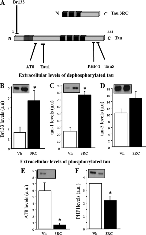
Extracellular tau promotes the release of intracellular tau, which is dephosphorylated in the extracellular medium. A, schematic representation of tau species and antibodies used in these experiments is shown. Black blocks represent the tubulin binding repeats; gray blocks represent exons 2 and 3. BR133 antibody recognizes the N terminus of tau, and Tau-1 antibody recognizes unphosphorylated Ser198, Ser199, Ser202. AT8 antibody recognizes tau when phosphorylated at Ser202 and Thr205. These antibodies recognize endogenous tau, but not tau3RC. Tau-5 is a phosphate-independent antibody that recognizes both endogenous tau and tau3RC, whereas PHF1 antibody requires phosphorylated tau at Ser396, Ser404 residues (17). B–D, culture medium from cells treated with vehicle solution (Vh) or with 1 μm tau3RC for 48 h was collected, and extracellular tau levels were measured by Western blotting using Br133 (B), Tau-1 (C), and Tau-5 (D) antibodies. Graphs represent the mean ± S.D. (error bars) from three individual experiments. *, p < 0.05. E and F, levels of extracellular phosphorylated tau were also measured by Western blotting using antibodies that specifically recognized the phosphorylated epitopes of tau protein, AT8 (E) and PHF1 (F). Blots were scanned to determine the proportion of phosphorylated tau. Graphs represent the mean ± S.D. from three individual experiments. a.u., arbitrary units. *, p < 0.05.

