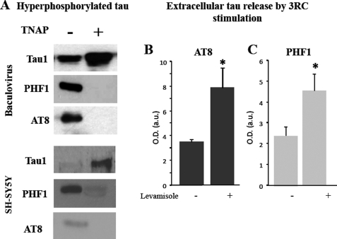
FIGURE 4.
TNAP dephosphorylates phospho-tau. A, several samples of hyperphosphorylated tau obtained from an insect cell culture infected with baculovirus expressing tau protein (upper panel) or phospho intracellular tau protein from SY5Y cells (lower panel) were incubated in the presence (+) or absence (−) of TNAP for 1 h at 37 °C. Phosphorylation degree of those samples were analyzed by Western blotting using Tau-1, PHF1, and AT8 antibodies. B and C, phosphorylation levels of endogenous tau released from tau3RC-treated cells (3RC; 1 μm for 48 h) in the presence or absence of 1 mm levamisole are shown. Culture media were collected, and extracellular tau levels were measured by dot-blotting using AT8 (B) and PHF1 (C) antibodies. Graph represents the mean ± S.D. (error bars) from three individual experiments. *, p < 0.05.

