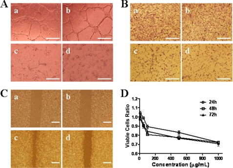
FIGURE 2.
WSS25 impaired the tube formation of HMEC-1 cells on Matrigel and migration. A, WSS25 inhibited the tube formation of HMEC-1 cells on Matrigel. HMEC-1 cells (90 μl) treated with WSS25 (10 μl) at different final concentrations (panel b, 6.25 μg/ml; panel c, 25 μg/ml; panel d, 50 μg/ml); or vehicle (panel a) were seeded into the 96-well plate precoated with 50 μl Matrigel for 10 h. B, WSS25 impaired the migration of HMEC-1 cells in a trans-well migration assay. HMEC-1 cells were seeded into the inner chamber with MCDB131 medium containing 10 μg/ml (panel b), 100 μg/ml (panel c), 1 mg/ml WSS25 (panel d) or vehicle (panel a). C, WSS25 inhibited the migration of HMEC-1 cells in a wound healing assay (panels a and c, control; panels b and d, 25 μg/ml). D, HMEC-1 cells were seeded into the 96-well plate. After 24 h of incubation, WSS25 was added to the final concentrations of 1 μg/ml, 10 μg/ml, 100 μg/ml, 500 μg/ml, or 1 mg/ml. The cell viabilities were determined by the MTT assay 24 h (○), 48 h (□), or 72 h ([tric]) later. The results are representative of triplicate experiments.

