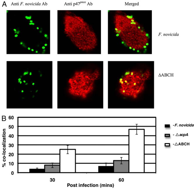
FIGURE 5.

Colocalization of the F. novicida WT strain and mutant strains with p47phox in macrophages. A, Colocalization of the F. novicida WT and ΔABCH or ΔacpA mutant strains with p47phox was determined at 30 and 60 min post-infection in MDMs. Francisella was detected following staining with goat anti-mouse Alexa Fluor 488 (green color), and p47phox was detected following staining with donkey anti-rabbit Alexa Fluor 546 (red color). Representative confocal microscopy images of F. novicida and ΔABCH colocalized with p47phox within MDMs are shown 60 min postinfection. The images are representative of 1000 infected cells examined from triplicate coverslips in three independent experiments. Original magnification ×63. B, Colocalization of the F. novicida WT, ΔacpA, and ΔABCH mutant strains with p47phox was quantified at 30 and 60 min postinfection. Analyses were based on examination of 1000 infected cells examined from triplicate coverslips in three independent experiments. The results shown are cumulative data of three experiments (mean ± SD of nine samples; triplicate samples were included in each test group in each experiment).
