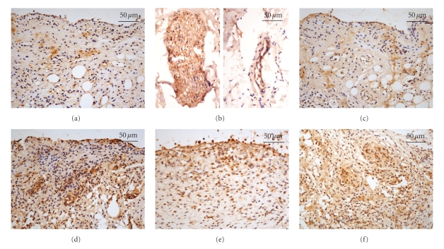
Figure 6.
Immunostaining for VIP in the synovial tissue. Histological analysis of the synovial tissue from the saline group shows weak staining for VIP in synovial cells (a) and moderate staining for VIP in endothelium, smooth muscle, and nerve fibers (b). (c) Synovial cells from the saline-EA group display weak immunoreaction for VIP. (d) Moderate to strong immunostaining for VIP is seen in the synovial tissue from the FCA-injected animals. An enhancement of the VIP immunoreactivity in the synovial tissue is observed in the FCA-EA group (e) but not in the FCA-EA (nonacupoint) group (f). Original magnification, 400x.

