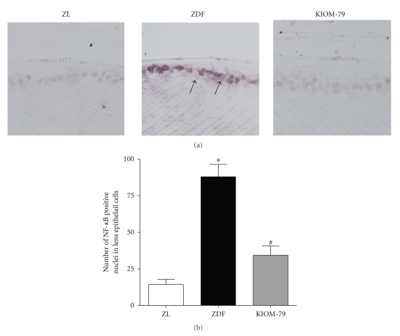
Figure 5.
Distribution of NF-κB in LECs detected by southwestern histochemistry. (a) Representative photomicrograph of lens from a normal Zucker lean rat (ZL), vehicle-treated ZDF rat (ZDF), and ZDF rat treated with KIOM-79 (KIOM-79). Positive signals (arrow) for activated NF-κB were mainly detected in nuclei of LECs of the vehicle-treated ZDF rat. X400 magnification. (b) Quantitative analysis of positive cells in LECs. Values in the bar graphs represent means ± SE, n = 8. *P < .01 versus normal ZL rats, # P < .01 versus vehicle-treated ZDF rats.

