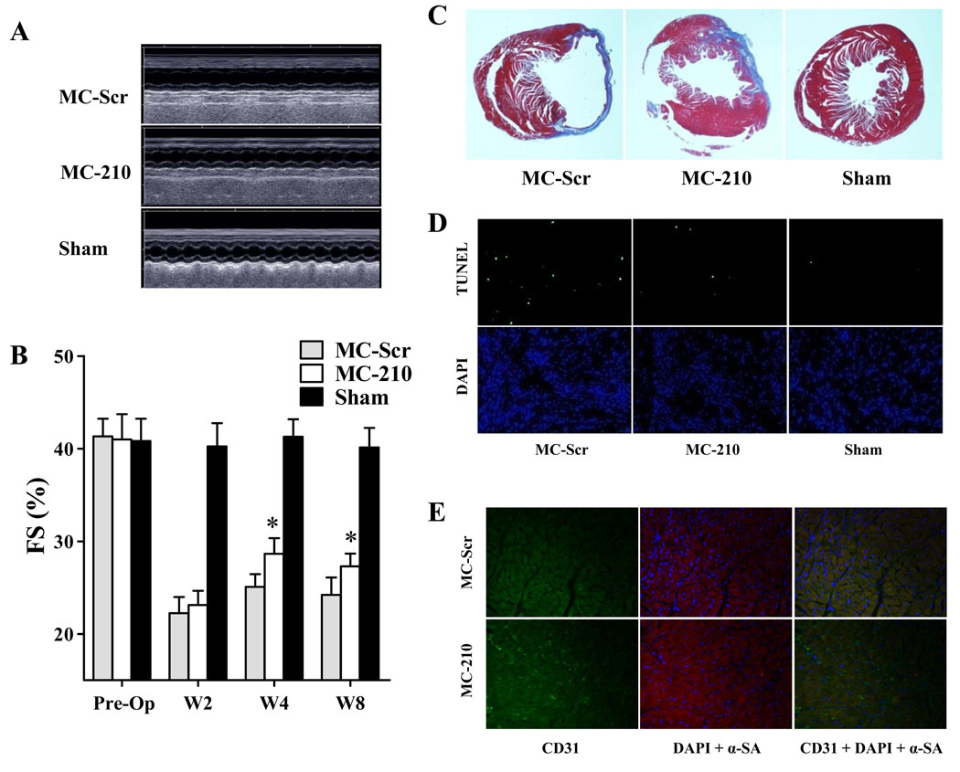
Figure 4.
Evaluation of cardiac function following MI after miR-210 treatment. (A) Representative echocardiogram of mice with LAD ligation after injection of MC-210, MC-Scr, or sham group at week 8. (B) Quantitative analysis of left ventricular fractional shortening (FS) among the 3 groups. Compared to MC-Scr control, animals injected with MC-210 had significant improvements in FS values at both week 4 and week 8. 2-way ANOVA was used for statistical analysis. (C) Representative Masson trichrome staining of explanted heart at week 8 showed increased wall thickness for the MC-210 group, confirming the positive functional imaging data seen in echocardiography. (D) TUNEL staining of explanted heart demonstrated significantly reduced apoptotic cells in MC-210 group compared to MC-Scr control group. (E) Immunofluorescence staining of CD31 endothelial marker (green) demonstrated increased neovascularization in the myocardium after MC-210 delivery compared to MC-Scr control. Cardiomyocyte staining is identified by α-sarcomeric actin (red) and nuclear staining is identified by DAPI (blue).

