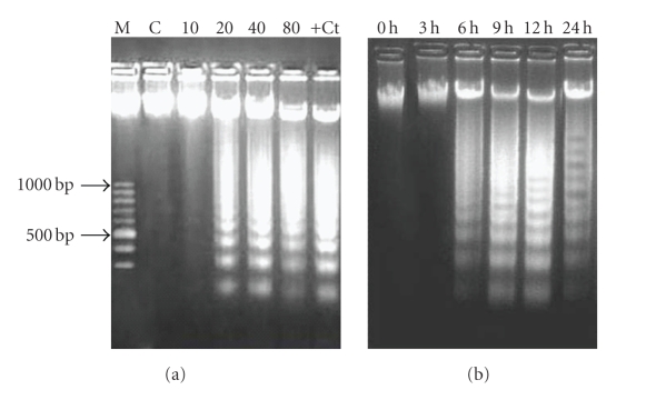
Figure 4.
Dose-dependent and time-dependent induction of DNA fragmentation in HL-60 cells after ZMS treatment. (a) 2 × 10⁶ HL-60 cells treated with different concentrations of ZMS extract for 24 hours. DNA was electrophoresed on 1.8% agarose gel and stained with ethidium bromide. M-Marker; C-Untreated HL-60 cells; 10-10 μg/mL of ZMS treated HL-60 cells; 20-20 μg/mL of ZMS treated HL-60 cells; 40-40 μg/mL of ZMS treated HL-60 cells; 80-80 μg/mL of ZMS treated HL-60 cells; +Ct− Camptothecin treated HL-60 cells. (b) 2 × 10⁶ HL-60 cells were treated with 20 μg/mL of ZMS extract and were incubated for 3 hours, 6 hours, 9 hours, 12 hours, and 24 hours. DNA was electrophoresed at respective time interval on 1.8% Agarose gel and stained with ethidium bromide.

