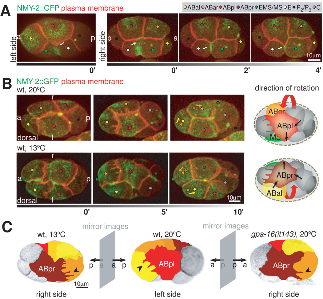
Figure 5. Handedness of Chiral Morphogenesis.
A. LR asymmetry of EMS cytokinesis correlates with asymmetry in ABpl/pr ventral protrusions. 3-D projection stills, left and right side views, respectively, with plasma membrane (red) and NMY-2 (green) fluorescently labeled. 0’ frames of left and right sides show the same point in developmental time in two carefully staged embryos. Arrows point at the contacts between the coalescing furrow of EMS and the ventral protrusions of ABpl and ABpr (0’) and at the developing midbody on the right side (2’ and 4’). B. Reversal of chiral morphogenesis by raising embryos at low temperature. Left panel: 3D- projection stills, dorsal views, right (r) and left (l) sides are indicated. Fluorescent markers are plasma membrane (red) and NMY-2 (green). Arrows in the last timepoint indicate the translocation of the corresponding cells, where the dots with lighter shade indicate the starting positions of the corresponding cells as shown in the first timepoint. Right panel: Schematic representation of wild-type and mirror-symmetric chiral morphogenesis. The three cells that perform the collective rotation are highlighted, red arrows indicate the handedness of the rotation. Black arrows indicate directions of force as deduced from the directionality of protrusions and cell movement. See also Movie S7. C. Overview of the effect of cold-treatment and gpa-16(it143) on chiral morphogenesis. 3D-projection stills from embryos expressing fluorescently labeled plasma membranes. Individual blastomeres are highlighted by false color representation to highlight mirror symmetry to the wild type; right side views of an embryo raised at 13°C and a gpa-16(it143) embryo at restrictive temperature (20°C) and left side view of a wild-type embryo. See also Movie S8.

