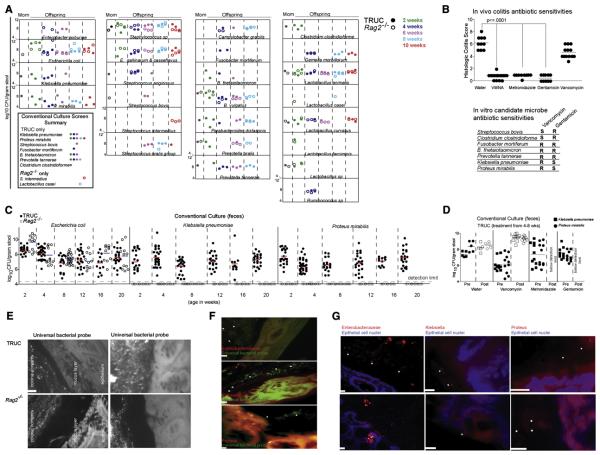
Fig 2. The presence of Klebsiella pneumoniae and Proteus mirabilis correlates with the presence of colitis in TRUC mice.
(A) Culture-based identification of bacterial species present in fecal samples from the same animals and time points as analyzed in Fig. 1. Species observed in more than one animal or in one animal at more than one time point are shown. Inset is a summary of bacterial species-level differences in the fecal microbiota of TRUC versus Rag2−/− mice observed in 2A. (B) Upper panel: Histologic colitis scores demonstrate the in vivo antibiotic sensitivities of TRUC colitis. Each dot represents an individual mouse treated for four weeks with the indicated antibiotics dissolved in drinking water. VMNA refers to treatment with a combination of vancomycin, metronidazole, neomycin, and ampicillin. Horizontal bars represent the mean. p-value ≤0.0001 defined by Mann-Whitney test. Lower panel: Summary of in vitro antibiotic sensitivities for several species selectively detected in TRUC fecal microbiota. (C) Culture-based survey of Gram-negative aerobes present in fecal samples from TRUC (shaded circles) and Rag2−/− (open circles) at 2-20 weeks of age. Fecal samples were collected and cultured twice at each time point from Rag2−/− mice. (D) In vivo sensitivity of Klebsiella pneumoniae (squares) and Proteus mirabilis (circles), as defined by culture-base surveys of TRUC fecal samples collected 1d before (shaded symbol) and 1d after (open symbol) treatment with antibiotic. Each dot represents data from a fecal sample obtained from one mouse. Horizontal bars represent the mean value. (E) Fluorescence In Situ Hybridization using an Oregon-Green® 488 conjugated “Universal bacterial” 16S rRNA-directed oligonucleotide probe (EUB 338) demonstrates the presence of bacteria in the mucus layer and directly adjacent to the epithelium in TRUC mice. Upper panels: TRUC, lower panels: Rag2−/−. A 10 micron scale bar for the panel is shown in the lower left of the first image. (F) Enterobacteriaceae (red), Klebsiella (red), and Proteus (red) were visualized adjacent to the epithelium in TRUC mice using fluor-conjugated 16S rRNA or 23S rRNA oligonucleotide probes [(pB-00914 (Enterobacteriaceae), pB-00352 (Klebsiella pneumoniae), pB-02110 (Proteus mirabilis)]. Sections were also hybridized with the EUB338 universal bacterial probe (green). Scale bars (10 micron) are shown for each image. (G) Enterobacteriaceae (red), Klebsiella (red), and Proteus (red) probe signals are seen adjacent to or along the epithelium in TRUC mice. Epithelial cell nuclei were stained with DAPI. White star symbols mark bacteria in (F) and (G). Scale bars (10 micron) are shown for each image.

