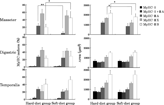
Fig. 4.
The proportions (%) and mean cross-sectional areas (μm2) of the different fiber types in the superficial masseter, anterior belly of digastric and anterior temporalis muscles. Values are means + SD. Significant differences between both groups: *P < 0.05; **P < 0.01. MyHC, myosin heavy chain.

