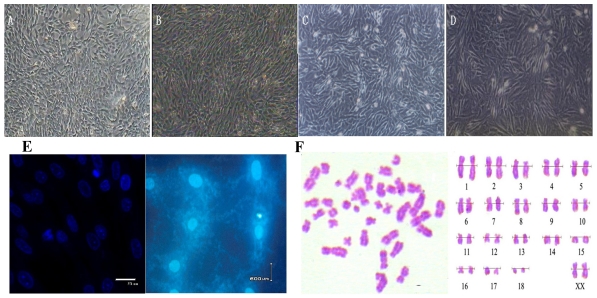
Figure 1.
Morphology, Mycoplasma contamination and karyotype of Siberian tiger cell line. (A) Primary cells (×100), the cells were typical long spindle-shape. (B) Subcultured cells (×100). (C) Cells before cryopreservation (×100). (D) Cells after recovery (×100). (E) Mycoplasma contamination Stained with Hoechst33258 and Positive control of Mycoplasma contamination; (F) Chromosome at metaphase (left) and karyotype (right) (×1,000).

